
- Sleep is an active biological state, not a period when the brain and body simply shut down.
- Sleep alternates between non-rapid eye movement (NREM) and rapid eye movement (REM) stages throughout the night.
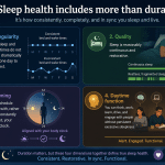
- Circadian timing and accumulated sleep pressure work together to regulate sleep and wakefulness.
- Brain activity, muscle tone, breathing, circulation, hormones, metabolism, temperature, and immune activity change during sleep.
- Deep NREM sleep is more prominent earlier in the night, while REM periods generally lengthen toward morning.
- Sleep architecture varies with age, health, medication, stress, substance use, schedules, and environmental conditions.
- Adequate, continuous sleep supports attention, learning, emotional regulation, metabolic function, and physical recovery.
What the physiology of sleep involves
The physiology of sleep describes the coordinated biological changes that occur as the body moves from wakefulness into sleep and back again.
The brain remains active during sleep, but its patterns of electrical activity, chemical signaling, communication, and responsiveness change. The cardiovascular, respiratory, endocrine, immune, metabolic, and temperature-regulation systems also adjust their activity.
Sleep is therefore an organized physiological state rather than an interruption of normal function.
No single theory fully explains why sleep is necessary. Energy regulation, cellular maintenance, immune coordination, memory processing, neural plasticity, and protection during vulnerable hours may all contribute. Sleep is best understood as a collection of overlapping processes rather than one restorative mechanism.
How the brain regulates sleep and wakefulness
Sleep and wakefulness emerge from connected networks in the hypothalamus, brainstem, basal forebrain, thalamus, and cerebral cortex.
Wake-promoting systems maintain alertness and responsiveness. As sleep begins, sleep-promoting neurons inhibit parts of these arousal networks. The hypothalamus helps connect sleep with circadian timing, body temperature, appetite, hormones, and autonomic regulation.
The brainstem contributes to arousal, breathing, muscle tone, and REM sleep. The thalamus changes how sensory information reaches the cerebral cortex, helping reduce conscious awareness of the environment.
There is no single brain structure that acts as a simple sleep switch. Different neural circuits coordinate the timing, depth, muscle activity, dreaming, and physiological features of sleep.
How the sleep–wake cycle is controlled
Two interacting systems exert much of the control over sleep:
- Circadian timing, which helps determine when the body is biologically prepared for sleep or alertness.
- Homeostatic sleep pressure, which generally increases during wakefulness and decreases during sleep.
Sleep is usually easiest when circadian timing and accumulated sleep pressure both support it.
Circadian rhythms and the internal clock
Circadian rhythms are approximately 24-hour patterns affecting sleep, alertness, body temperature, hormone release, digestion, and metabolism.
Light is the strongest environmental timing signal. Meals, physical activity, work schedules, and social routines also help synchronize rhythms throughout the body.
The circadian system does not produce sleepiness at one fixed hour. Instead, it creates changing periods of stronger biological support for sleep and wakefulness. This helps explain why a person can experience an evening “second wind” despite having been awake for many hours.
The role of the suprachiasmatic nucleus
The suprachiasmatic nucleus, or SCN, is a small group of cells in the hypothalamus that acts as the brain’s central circadian timekeeper.
It receives information about environmental light from the eyes and helps coordinate daily patterns of sleep, melatonin release, body temperature, and other physiological functions.
Organs and tissues also contain peripheral clocks. Irregular sleep, meal, and activity schedules can therefore create internal misalignment even when total sleep duration appears adequate.
Melatonin and light exposure
Melatonin is released by the pineal gland as biological night approaches. It helps communicate circadian timing but does not independently force the body to sleep.
Evening light can suppress melatonin and delay its onset. The effect depends on the timing, intensity, duration, and wavelength of the light exposure. Controlled research found that exposure to ordinary room light before bedtime delayed melatonin onset and shortened the duration of melatonin production compared with dim light. [1]
Reducing bright evening light and obtaining outdoor light after waking can provide clearer timing signals. Screens are one source of evening light, but their effects also depend on brightness, viewing duration, distance, content, and the surrounding environment.
Sleep pressure and adenosine
Sleep pressure generally becomes stronger the longer a person remains awake. Adenosine, a substance associated with cellular energy use, contributes to this process.
Caffeine promotes alertness primarily by blocking adenosine receptors rather than eliminating the need for sleep.
Sleep reduces homeostatic pressure, but it is misleading to claim that deep sleep simply “clears” adenosine. Sleep pressure reflects several interacting biological processes and cannot be reduced to the removal of one chemical.
Circadian alerting signals can temporarily counteract high sleep pressure. This is why exhaustion does not always result in immediate sleep and why sleeping at an unusual biological time may be difficult.
Understanding the stages of sleep
Sleep is divided into NREM and REM sleep. NREM includes three stages that progress from light sleep to deep slow-wave sleep.
These stages recur in cycles throughout the night. A cycle is often described as lasting about 90 minutes, but its duration and composition vary between individuals and across the same night.
NREM stage 1
N1 is the transition between wakefulness and sleep. Brain activity begins to slow, muscles relax, and awareness of the surroundings becomes less dependable.
Brief muscle jerks or a sensation of falling may occur. Because sleep remains light, the person may awaken easily and may not realize that sleep occurred.
NREM stage 2
N2 is a more stable stage of sleep. It includes bursts of electrical activity called sleep spindles and larger patterns called K-complexes.
These features help regulate sensory responsiveness and are associated with aspects of memory processing. Heart rate and breathing settle, muscles relax further, and body temperature continues to decline.
N2 generally occupies a substantial proportion of an adult’s sleep period.
NREM stage 3
N3 is deep sleep, also called slow-wave sleep. It is characterized by prominent slow-wave brain activity and reduced responsiveness to the environment.
Heart rate and breathing are generally slower and more regular than during wakefulness or REM sleep. Waking suddenly from N3 can produce temporary confusion and impaired performance known as sleep inertia.
Deep sleep is usually concentrated in the earlier part of the night, when homeostatic sleep pressure is strongest.
N3 is associated with physiological recovery and some forms of memory processing, but it should not be presented as the only restorative stage. Recovery depends on the duration, continuity, timing, and overall architecture of sleep.
REM sleep
REM sleep includes rapid eye movements, active brain patterns, vivid dreaming for many people, variable breathing and heart rate, and a substantial reduction in skeletal-muscle activity.
Dreaming can also occur during NREM sleep, so dreaming is not exclusive to REM.
During REM, the nervous system strongly inhibits most skeletal muscles. This temporary loss of muscle tone is called REM atonia. It limits large body movements while dream-related brain activity is occurring, although breathing muscles, eye muscles, and some small muscle groups remain active.
REM atonia should not be described as rebuilding muscles, decompressing joints, reducing inflammation, or improving flexibility. Those are not established functions of the phenomenon.
REM periods generally become longer toward morning. Repeatedly shortening the final part of the sleep period may therefore reduce REM sleep disproportionately.
How brain activity changes during sleep
The sleeping brain does not become uniformly inactive. Electrical rhythms and communication between brain regions change from stage to stage.
During NREM sleep, cortical activity becomes progressively slower and more synchronized. REM sleep produces patterns that resemble wakefulness in some brain regions even though the person remains asleep and largely disconnected from the environment.
These patterns can be measured using electroencephalography, or EEG. EEG recordings help identify sleep stages and transitions, but they do not provide a single measurement of how restorative someone’s sleep was.
Neurotransmitters and arousal
Wakefulness and sleep depend on changes in several chemical systems.
Acetylcholine, norepinephrine, serotonin, histamine, dopamine, orexin, GABA, and adenosine all contribute, but they do not follow one uniform pattern throughout the night.
Wake-promoting systems help sustain attention and responsiveness. Sleep-promoting inhibitory signaling suppresses parts of those systems during NREM sleep. REM sleep has a distinct chemical profile that contributes to its combination of active brain patterns, dreaming, and reduced muscle tone.
Sensory processing
The sleeping brain filters much of the sensory information arriving from the environment, but it does not become completely isolated.
Predictable background sounds may be ignored, while an alarm, a baby’s cry, pain, breathing difficulty, or an unfamiliar noise can trigger arousal. This selective responsiveness allows sleep to continue while preserving some ability to react to important conditions.
What the body does during sleep
Sleep involves coordinated changes in circulation, respiration, hormones, metabolism, temperature, and immune activity.
These changes are not constant throughout the night. They vary with sleep stage, circadian phase, health, medication, breathing, and brief awakenings.
Heart rate, blood pressure, and breathing
Heart rate, blood pressure, and sympathetic nervous system activity commonly decline during stable NREM sleep. REM sleep is generally more variable and can include temporary cardiovascular increases.
Breathing usually becomes slower and more regular during NREM sleep. During REM, breathing may become less regular because of changes in brain activity and muscle control.
Brief arousals can produce short surges in heart rate and blood pressure even when the person does not remember waking. Experimental research found that repeated sleep fragmentation delayed the normal sleep-related reduction in blood pressure. [2]
Persistent loud snoring, gasping, choking, witnessed breathing pauses, morning headaches, or substantial daytime sleepiness may indicate a sleep-related breathing disorder and should be discussed with a healthcare professional.
Hormonal regulation and recovery
Sleep influences hormones involved in growth, stress, reproduction, appetite, and metabolism.
Growth hormone secretion is commonly associated with sleep onset and early-night slow-wave sleep. However, the relationship is not absolute, and growth hormone release may occur outside slow-wave sleep.
Cortisol follows a strong circadian rhythm. Levels are commonly lower during the early biological night and begin rising before waking. Sleep can influence the timing of this rise, but cortisol should not be described as remaining uniformly suppressed throughout the night. [3]
Sleep supports physiological conditions associated with tissue maintenance and recovery. This does not mean every injury heals overnight or that one sleep stage independently performs all tissue repair.
Appetite hormones
Sleep can influence hormones involved in appetite, but the relationship is more complicated than the common claim that insufficient sleep always lowers leptin and raises ghrelin.
One controlled study of 12 healthy young men found lower leptin, higher ghrelin, and increased hunger after restricted sleep. [4] However, other studies have not consistently reproduced both hormonal changes.
Appetite during sleep loss may also be affected by stress, reward processing, fatigue, food availability, meal timing, and the additional time available for eating.
Metabolism and energy balance
Sleep and circadian timing influence glucose regulation, insulin sensitivity, appetite, digestion, food choices, and energy use.
In a randomized crossover study, five nights with four hours in bed reduced whole-body and peripheral insulin sensitivity in healthy participants compared with five nights with eight hours in bed. [5]
The size and clinical importance of such effects depend on the severity and duration of sleep restriction, the population studied, diet, activity, health, and experimental conditions.
Adequate sleep supports metabolic regulation, but sleep alone is not a treatment for diabetes, obesity, or another metabolic disorder.
Immune activity and inflammation
Sleep and immunity communicate in both directions. Immune signaling can increase fatigue and sleepiness during illness, while sleep affects the timing and activity of immune responses.
Experimental sleep restriction has altered cytokine production and T-helper-cell signaling in human participants. These findings indicate that insufficient sleep can change immune regulation rather than simply “weakening” or “strengthening” the immune system in one uniform way. [6]
Immune effects vary with sleep duration, circadian timing, infection, stress, age, medication, and the specific immune response being measured.
Temperature regulation
Core body temperature usually declines as biological night develops and remains lower during much of the sleep period. Heat loss through the skin helps support this reduction.
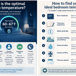
An environment that is uncomfortably warm or cold may delay sleep or cause brief arousals. A cool, dark, and quiet bedroom can support sleep, although environmental adjustments cannot correct every medical or circadian cause of disrupted sleep.
Why sleep architecture matters
Sleep architecture refers to the timing, order, duration, and continuity of NREM and REM stages.
A typical sleep period begins with NREM sleep and progresses into REM before the sequence repeats. Deep NREM sleep is concentrated more heavily in the early part of the night, while REM becomes more prominent later.
This progression is not mechanical. Healthy people may briefly awaken between cycles, and sleep architecture varies from night to night.
Total sleep duration is important, but it does not reveal whether sleep was appropriately timed, continuous, or repeatedly disrupted.
How age affects sleep
Infants spend more time asleep and have sleep patterns that differ substantially from those of adults. Sleep generally becomes more consolidated through childhood.
In later adulthood, deep sleep may become less abundant and awakenings may become more frequent. Age is only one influence. Health conditions, medication, pain, activity, mood, alcohol use, and living circumstances can alter sleep at any age.
Factors that can change sleep stages
Common influences include:
- Prior sleep loss
- Irregular schedules and shift work
- Evening and nighttime light exposure
- Alcohol, caffeine, medications, and other substances
- Stress, anxiety, pain, and illness
- Sleep-related breathing or movement disorders
- Noise, temperature, and other environmental disturbances
These factors often interact. Stress may delay sleep, for example, while an irregular schedule places sleep at an unfavorable circadian time.
Altered sleep stages alone do not establish a diagnosis. Sleep data must be interpreted alongside symptoms, schedule, medical history, and the quality of the measurement.
Fragmented sleep and health
Fragmented sleep occurs when sleep is repeatedly interrupted or shifted into lighter stages.
Possible causes include:
- Environmental noise
- Pain
- Acid reflux
- Stress or anxiety
- Breathing difficulties
- Movement disorders
- Alcohol or other substances
- Medication effects
- An uncomfortable sleep environment
Brief awakenings are normal and are often forgotten. They become more concerning when they are frequent, prolonged, or accompanied by daytime impairment.
Persistent fragmentation combined with excessive daytime sleepiness, morning headaches, impaired concentration, mood changes, breathing pauses, or unusual movements during sleep may warrant medical evaluation.
How sleep supports memory and brain function
Sleep provides conditions in which recently acquired information can be stabilized, reorganized, and integrated with existing knowledge.
NREM sleep is associated with the reactivation and stabilization of some forms of learning. REM sleep may contribute to emotional and associative memory processes. These roles overlap, however, and research does not support assigning each type of memory exclusively to one stage.
Recent experimental work suggests that slow-wave and REM sleep can make complementary contributions to emotional-memory processing, while also showing that stage-specific effects are more complicated than earlier theories proposed. [7]
Sleep does not replace attention, understanding, or practice. It helps create physiological conditions in which learning can be retained and integrated.
Effects of insufficient sleep
Insufficient sleep can impair:
- Attention
- Reaction time
- Working memory
- Judgment
- Emotional regulation
- Learning
- Decision-making
In a controlled experiment, participants assigned four or six hours in bed per night for 14 nights developed cumulative, dose-dependent performance deficits. Their subjective sleepiness did not fully reflect the extent of their cognitive decline. [8]
People may therefore feel that they have adapted to restricted sleep even while objective performance continues to deteriorate.
Brain fluid movement and the glymphatic system
The brain has fluid-transport pathways involved in moving substances through and around brain tissue. These pathways are often discussed under the term glymphatic system.
Animal research indicates that brain state, neural activity, cerebrospinal fluid movement, and molecular transport are connected. However, the popular claim that deep sleep definitively “washes toxins” from the human brain presents an unsettled area of research as established fact.
A 2024 mouse study found that synchronized neuronal activity during sleep could drive ionic waves associated with cerebrospinal fluid movement and molecular clearance. [9]
Another 2024 mouse study, using a different measurement method, found that the clearance of a fluorescent molecule was reduced during sleep and anesthesia. [10]
These studies examined different processes and used different methods, which may partly explain their apparently conflicting conclusions. The evidence supports saying that sleep affects brain fluid dynamics. It does not yet justify presenting nightly “brain cleaning” as a simple, proven human mechanism.
Conclusion
Sleep is a coordinated interaction between the brain, body, internal clock, and accumulated need for sleep.
Across NREM and REM cycles, brain activity, muscle tone, breathing, circulation, hormones, metabolism, temperature, immune signaling, memory processing, and sensory responsiveness all change.
No single stage or mechanism accounts for every benefit of sleep. Sleep health depends on sufficient duration, appropriate timing, regularity, continuity, and the absence of untreated disorders.
Understanding this physiology makes sleep feel less like lost time and more like an active biological process that supports functioning throughout the following day.
Frequently asked questions
What is the physiology of sleep?
It is the study of the biological changes that occur during sleep, including changes in brain activity, breathing, circulation, hormones, metabolism, temperature, sensory processing, and muscle tone.
What are the two main types of sleep?
The two main types are NREM and REM sleep. NREM is divided into stages N1, N2, and N3.
What controls the sleep–wake cycle?
Circadian timing interacts with homeostatic sleep pressure. Circadian rhythms provide time-of-day information, while sleep pressure generally increases during wakefulness and decreases during sleep.
What does melatonin do?
Melatonin helps signal biological night and contributes to circadian timing. It does not act as an on-off switch that independently forces sleep.
Why is deep sleep important?
Deep NREM sleep is associated with slow-wave brain activity, reduced environmental responsiveness, physiological recovery, and some forms of memory processing. It is one part of healthy sleep architecture rather than the only restorative stage.
What is REM atonia?
REM atonia is the strong reduction of most skeletal-muscle activity during REM sleep. It limits large movements while the brain is producing internally generated dream experiences.
Does sleep remove waste from the brain?
Sleep affects brain fluid movement, but the mechanisms and net effects on molecular clearance remain disputed. Current evidence does not support reducing the process to the claim that sleep simply washes toxins from the human brain.
How does insufficient sleep affect metabolism?
Sleep restriction can interfere with insulin sensitivity, appetite, eating behavior, and glucose regulation. The effects involve several physiological and behavioral pathways rather than one pair of appetite hormones.
When should sleep problems receive medical attention?
Medical advice may be appropriate when sleep problems persist or involve substantial daytime sleepiness, witnessed breathing pauses, gasping, morning headaches, dangerous drowsiness, frequent awakenings, or unusual movements and behaviors during sleep.
References
- Gooley JJ, Chamberlain K, Smith KA, et al. Exposure to room light before bedtime suppresses melatonin onset and shortens melatonin duration in humans. Journal of Clinical Endocrinology & Metabolism. 2011;96(3):E463–E472. DOI: 10.1210/jc.2010-2098. (pubmed.ncbi.nlm.nih.gov)
- Carrington MJ, Trinder J. Blood pressure and heart rate during continuous experimental sleep fragmentation in healthy adults. Sleep. 2008;31(12):1701–1712. DOI: 10.1093/sleep/31.12.1701. (pubmed.ncbi.nlm.nih.gov)
- Davidson JR, Moldofsky H, Lue FA. Growth hormone and cortisol secretion in relation to sleep and wakefulness. Journal of Psychiatry & Neuroscience. 1991;16(2):96–102. PMID: 1911740. (pmc.ncbi.nlm.nih.gov)
- Spiegel K, Tasali E, Penev P, Van Cauter E. Brief communication: Sleep curtailment in healthy young men is associated with decreased leptin levels, elevated ghrelin levels, and increased hunger and appetite. Annals of Internal Medicine. 2004;141(11):846–850. PMID: 15583226. (pubmed.ncbi.nlm.nih.gov)
- Rao MN, Neylan TC, Grunfeld C, et al. Subchronic sleep restriction causes tissue-specific insulin resistance. Journal of Clinical Endocrinology & Metabolism. 2015;100(4):1664–1671. DOI: 10.1210/jc.2014-3911. (pubmed.ncbi.nlm.nih.gov)
- Axelsson J, Rehman JU, Åkerstedt T, et al. Effects of sustained sleep restriction on mitogen-stimulated cytokines, chemokines and T helper 1/T helper 2 balance in humans. PLOS ONE. 2013;8(12):e82291. DOI: 10.1371/journal.pone.0082291. (pmc.ncbi.nlm.nih.gov)
- Yuksel C, et al. Both slow wave and rapid eye movement sleep contribute to emotional memory consolidation. Communications Biology. 2025. DOI: 10.1038/s42003-025-07868-5. (pubmed.ncbi.nlm.nih.gov)
- Van Dongen HPA, Maislin G, Mullington JM, Dinges DF. The cumulative cost of additional wakefulness: Dose-response effects on neurobehavioral functions and sleep physiology from chronic sleep restriction and total sleep deprivation. Sleep. 2003;26(2):117–126. DOI: 10.1093/sleep/26.2.117. (pubmed.ncbi.nlm.nih.gov)
- Jiang-Xie LF, Drieu A, Bhasiin K, et al. Neuronal dynamics direct cerebrospinal fluid perfusion and brain clearance. Nature. 2024;627:157–164. DOI: 10.1038/s41586-024-07108-6. (nature.com)
- Miao A, Luo T, Hsieh B, et al. Brain clearance is reduced during sleep and anesthesia. Nature Neuroscience. 2024;27:1046–1050. DOI: 10.1038/s41593-024-01638-y. (nature.com)